By Alexis Dobranowski
Sunnybrook’s radiation and clinical trials teams took a huge leap forward in February when they took the first human images on the Odette Cancer Centre’s new MR-Linac – the Elekta Unity.
The new technology is the first machine in the world to combine radiation and high-resolution magnetic resonance imaging (MRI), and will let doctors at the Odette Cancer Centre target tumours and monitor their response to radiation with unprecedented precision — even as a tumour moves inside the body — thanks to the machine’s real-time MRI guidance.
As the first Canadian centre to install an MR-Linac, Sunnybrook’s team is leading the way and helping to set up and conduct clinical trials that will establish the best treatment protocols for this new machine.
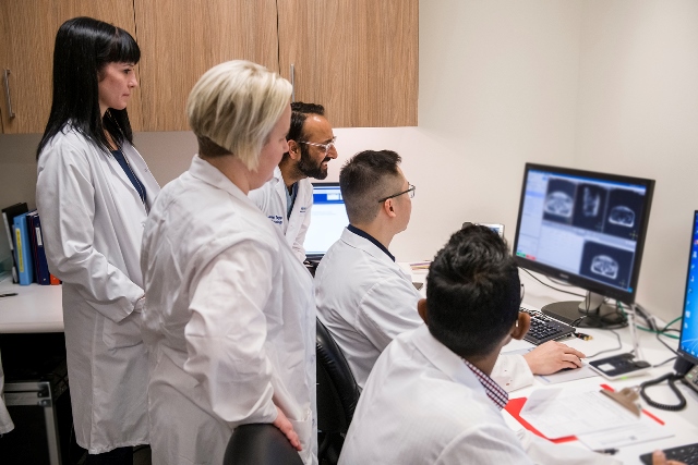
The first step is an imaging study that began in February. Study participants will have their imaging and treatment as usual, and then will undergo an extra MRI on this new machine. The imaging study aims to establish the MR (magnetic resonance) scan protocols that will be used to treat patients on the MR-Linac, explained Dr. Claire McCann, medical physicist and lead of the imaging study.
“We will use the images in our study to help us develop procedures to allow us to adapt a patient’s radiation treatment to changes in the tumour that may occur over time,” she said.
Currently, a patient has a CT image taken before radiation. That image is used to plan the patient’s treatment, like where to aim the radiation beam. For some patients, we also get a single MR image, which helps us with this radiation planning.
“With our new MR-Linac, we will be able to get MR images before every radiation treatment,” said radiation oncologist Dr. Arjun Sahgal, clinical director of the MR-Linac. “Those MR images will be used to help us ensure the most accurate treatment based on the tumours specific location each and every day. These images will also allow us to monitor how the tumour responds and determine the best way to adapt the treatment to those changes in real time.”
Images taken during the imaging study will help the team develop the clinical workflows – procedures and processes for how they manage any tumour changes, to ensure the most effective way to adapt the patient’s treatment.
Anatomical imaging lets the team see the tumour’s anatomy. Functional imaging looks at biological changes, for example changes in tumour blood flow as a result of radiation treatment.
“As our experience with this technology develops, we will add functional imaging to supplement the anatomical imaging, which will allow us to adapt radiation treatment not only to changes in tumour size, shape and location but based on biological response to the radiation as well,” Dr. McCann says.
Once the combined MR-Linac system receives full Health Canada approval, clinical trials will begin where patients will be treated with this technology using the imaging parameters and workflow developed as part of this imaging study for MR-guided adaptive radiotherapy.
Alexis Dobranowski is a Communications Advisor at Sunnybrook Health Sciences Centre.


