By Season Osborne
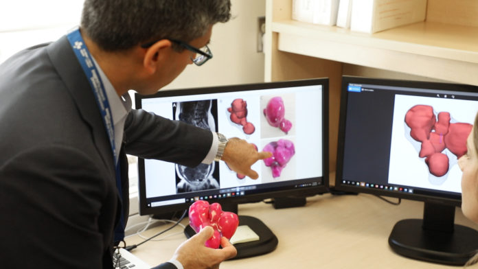
The uterus fits in the palm of Dr. Sony Singh’s hand. The large pink lumps inside the clear, plastic 3D-printed model are fibroids, or tumours, and there are more than 50 of them. To ensure his patient could carry a child in the future, Dr. Singh had to do something that had never been done before.
Maureen had suffered for years with abdominal pain. Over the past six years, she was told by five doctors that she had so many fibroids in her uterus, her only option was to have a hysterectomy – complete removal of her womb. She refused this option.
“I will die with my womb. Nobody will touch it,” says Maureen (who did not want her last name used).
She was referred to the Shirley E. Greenberg Women’s Health Centre at The Ottawa Hospital, where she saw the Minimally Invasive Gynecology team of doctors and nurses. Dr. Singh, a surgeon and the Elaine Jolly Research Chair in Surgical Gynecology, told Maureen he could remove all the fibroids, and she would not need a hysterectomy.

Dr. Teresa Flaxman, Research Associate at The Ottawa Hospital, said it was difficult to see tumours in the patient’s uterus on an MRI. So, she contacted the hospital’s 3D Printing Lab. With the opening of the lab in February 2017, the hospital became the first in Canada to have an integrated medical 3D-printing program for pre-surgical planning and education.
“3D printing is revolutionizing the way we look at anatomy,” said Orthopaedic Surgeon and Oncologist Dr. Joel Werier, who has used 3D-printed models of his patients’ hips and bones since the lab opened. “It adds another perspective to how we view tumours, how we plan our surgery techniques, and our ability to offer precision surgery.”
Bones are relatively easy to create from CT scans and MRIs, said Dr. Flaxman. However, soft tissues, such as uterine tissue, is harder to identify, and a model hadn’t been made of one before.
Dr. Flaxman and other researchers from the Women’s Health Centre worked with the lab to create 3D images from an MRI of Maureen’s uterus. Then the lab printed a model that allowed them to see exactly where the fibroids and the lining of the uterus were located. Dr. Singh successfully removed the fibroids, sparing Maureen from having a hysterectomy.
“This model helped to provide a good visual aspect,” said Dr. Singh. “To have a model in my hands during surgery was incredible.”
Dr. Adnan Sheikh, Director of The Ottawa Hospital’s 3D Printing Program said that, since this recent success, the lab is working on other similar projects with the Minimally Invasive Gynecology team to offer other women alternatives to major surgery in the future.
Season Osborne is the Publications Officer at The Ottawa Hospital Foundation.

