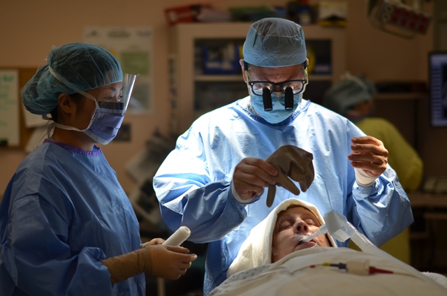
Fate led me to the field of neurosurgery. As a young man, I was a keen cyclist and skier. Then, when I was in medical school, my skiing ability began to deteriorate for no reason. From a loss of ability to ski, my walking declined and I eventually ended up in a wheelchair. It took some time but I was finally diagnosed with a spinal tumor in my lower back and had high-risk surgery that allowed me to get my life back. I figured that I would like to offer the same chance to others so I decided to become a neurosurgeon. Fraser Health recently shot a video of a procedure to remove a brain tumour from one of my patients, and tweeted the surgery and a Q&A with me. Here are my five easy steps to remove a brain tumor. But don’t try this at home.
Step 1: Diagnosis
Before you can remove a brain tumor, you have to make the diagnosis. This is the hardest part. Meningiomas, the most common benign brain tumors we see, are usually slow growing and may attain considerable sizes due to their ability to compress the surrounding brain in an insidious fashion. Malignant tumors may present more rapidly, with the development of weakness, or cognitive problems. Sometimes tumors can present during investigation of what may be an entirely unrelated complaint. For example, pituitary tumors either present with endocrine abnormalities (difficulty with conception, loss of libido, menstrual irregularities, weight loss or gain, or blood pressure problems) or visual disturbances.
MORE: THE WAR AGAINST SUPERBUGS
Step 2: Assembling the team
Just as it takes a village to raise a child, it takes a team to remove a brain tumor. The critical components include: surgeon, anaesthetist, scrub nurse(s), circulating nurses, OR assistants, anaesthesia assistants, and cleaners.
Step 3: Opening the head
Once the patient is asleep and properly positioned, their head is placed in a special device which prevents any movement (Mayfield 3 pin headrest). This allows the surgeon to make precise millimetre or even micrometre movements without fear. Most of us currently use a NeuroNavigational tool that allows precise localization of any lesion in the brain. This minimizes the size of the incision we make and can also reduce operative time. It is based on pre-operative imaging and the relationship of superficial anatomy of the scalp, face and skull relative to its contents.
Step 4: Removing the Tumour
This is the most delicate part of the procedure. The less we disturb the surrounding brain, the easier it is on the patient. Once we expose the tumor in its entirety, we can remove it as one specimen if it is small, or piecemeal if it is large. Depending on its adherence to the brain, this can take minutes or hours. Sometimes we use an Ultrasonic aspirator which liquefies the tumor while preserving blood vessels that run through it. This is often used for large meningiomas. The pathologist then attends the case and tests the tumor to determine its origin. Often we get an answer within 15 minutes as to the nature of the tumor (benign or malignant, primary or secondary).
MORE: DOCTORS RECONNECT WITH LOST BOY
Step 5: Closing the head
Once we know there is no bleeding, we can suture the dura together using fine stitches. We then replace the bone flap and fix it in place using special titanium rivets that hold the skull solid. Muscle and scalp layers are then sutured or stapled together, and a head dressing is applied. The patient is then reversed from anaesthesia and taken to the recovery room for observation for the next four to six hours. Once stable, they are taken to the ward where they remain under close observation. And that’s it.
Photos and video
Watch the entire surgery
View the photo gallery


