A Smarter Prostate Cancer Biopsy
Researchers at Sunnybrook Health Sciences Centre are perfecting a type of GPS navigation and roadmap for prostate cancer biopsies, by combining 3D MRI (multiparametric magnetic resonance imaging) and ultrasound to better guide the biopsy needle.
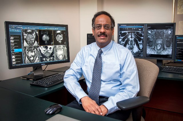
Standard biopsies use ultrasound alone which limits visualization of the tumour. With what radiologist Dr. Masoom Haider calls the ‘smart biopsy, men undergo an image-navigated biopsy to the area of cancer only, resulting in far fewer biopsies for the patient, and more accurate assessment of the tumour.
Studies conducted by Dr. Haider and Dr. Laurent Milot in Medical Imaging, and oncologists Drs. Laurence Klotz, Danny Vesprini, Andrew Loblaw and Robert Nam of Sunnybrook’s Odette Cancer Centre’s Genitourinary Cancer Care team show significant cancers can be found based on three to four needle biopsies or tissue samples using smart biopsy, compared to 12 needles with the standard procedure.
MORE: THE LATEST IN PERSONALIZED CANCER CARE
“With a clearer view, we are also detecting tumours that were missed on first biopsy done the standard way.” The three-dimensional view is made up of an MRI taken just before the procedure that is fused with ultrasound imaging recorded during the procedure. “MRI substantially improves our ability to identify and characterize tumours,” says Dr. Haider, chief of the Department of Medical Imaging at Sunnybrook, and a professor in the department of Medical Imaging at the University of Toronto, who helped refine the technologies to localize cancers with MRI.
Sunnybrook’s Odette Cancer Centre is one of only a few centres in Canada conducting research with a large volume of navigated smart biopsies.
Ultrasound for faster breast cancer treatment tracking
Knowing sooner helps better tailor treatment. That’s the premise behind new imaging research known as Quantitative Ultrasound being led by Dr. Gregory Czarnota. A radiation oncologist of the Odette Breast Cancer care team, Dr. Czarnota is using low-intensity ultrasound to detect cancer cell death from chemotherapy, in one to four weeks, in patients with locally advanced breast cancer.
“A faster determination of how a cancer is responding means clinicians can modify to a course of more effective treatment, essentially individualizing treatment that much sooner for the patient,” says Dr. Czarnota, who is also the head of Radiation Oncology at the Odette, and a senior scientist at Sunnybrook Research Institute.
Dr. Czarnota and his team use ultrasound as functional imaging to monitor tumour metabolic activity, applying special software to detect the absence or presence of cell death. This technology has been trialed with over 100 women with locally advanced breast cancer who had pre-surgery or neoadjuvant chemotherapy, a frequent treatment approach to shrink typically large breast tumours for better breast-conserving surgery.
“The results proved that the technology works – that within one to four weeks we can demonstrate whether a specific chemotherapy was going to work,” says Dr. Czarnota. Cancer response to treatment is usually only known after months of treatment and is determined by MRI or CT (computed tomography).
“We’re now at the stage where the technology is being expanded to potentially benefit other patients through more clinical trials in other centres,” he says.
Minimally invasive approach may replace liver biopsy
What if patients could avoid a biopsy and undergo an equally telling but non-invasive test? That is the question posed by Dr. Laurent Milot, an abdominal radiologist in Medical Imaging at Sunnybrook, who collaborates with the Gastrointestinal and Genitourinary Cancer Care teams at the Odette Cancer Centre.
MORE: IT’S TIME TO START USING THE M-WORD
Dr. Milot is the first in the world to show in cancer that MRI with a newer contrast agent, gadofosveset trisodium, better characterizes whether or not an abnormality is cancer, compared to MRI with standard gadolinium contrast agent.
“This technique could significantly benefit patients with spots on the liver that are highly suspicious of potential spread from colorectal cancer, especially as liver biopsy can be risky,” says Dr. Milot.
His research published in the Journal of Magnetic Resonance Imaging shows that after 10 minutes, MRI with gadofosveset trisodium produces images with a distinct black hollow or leakage of contrast agent in cancer lesions, compared to images of pooling of contrast agent in benign ones.
He is using this technique in early clinical trials with patients and collaborating with Odette oncologists to use the data for vital surgical planning.
To better characterize tumours, Dr. Milot, an affiliate scientist at Sunnybrook Research Institute who completed his Medical Imaging specialty at l’Université de Claude-Bernard Lyon 1 in France, is also researching the use of microscopic bubbles or microbubbles within blood vessels with contrast-enhanced ultrasound. Cancer lesions distinctly show rapid ‘wash out’ of microbubbles due to the abnormal vasculature of tumours, while benign lesions accumulate or retain them.

